The human heart is the vital organ in the body that keeps our body in circulation with its rhythmic (round-the-clock) supply of nutrients. On the other hand, others are of the opinion that the heart is likewise affected by the gradual onset of weakness and sickness. The main cardiovascular problem is, however, coronary disease which is not only widespread but also prevalent globally. Coronary Artery Disease results from the accumulation of plaques along the coronary arteries, making it harder for the oxygen-rich blood to be transported to the heart muscle. Artery plaque buildup can narrow the blood vessels, leading to reduction of blood flow to the heart. The resultant accumulation of such cardiovascular issues as chest pains or / discomfort, heart failure or arrhythmia, etc. worsens according to the severity of confinement.
Although innovative drugs, lifestyle change and careful systems made it possible to combat coronary disease, these saw knowledgeable development in the sector of imaging and diagnosis. Intravascular ultrasound (IVUS) is one of the basic restoration imaging methods in which physicians can visualise and understand the degree of the disease from the inside just like they would by looking at those arteries. Through this kind of data, specialists can make precise diagnoses and make appropriate treatment plans for patients. With its unique features, IVUS is a vital player in the management of coronary artery disease in today’s world. Let’s look at the evolution of this innovation in cardiology.
The clear view of Coronary Blood Vessels was witnessed
Coronary angiography, for a long period of time, has remained the golden standard of coronary artery disease assessment and therapy. However, this technique may have a few drawbacks. Angiograms simply make figure-of-speech about the scope of obstruction in veins. Therefore, we will not have an inside and out assessment with details like nature and plaque production, clear measurement of the lumen opening for instance, to avoid the segment being too long or too short, which are necessary for TIA management. In comparison to coronary angiography, which is constrained by the limit of imaging, IVUS erases that limit by visualising coronary vessels better.
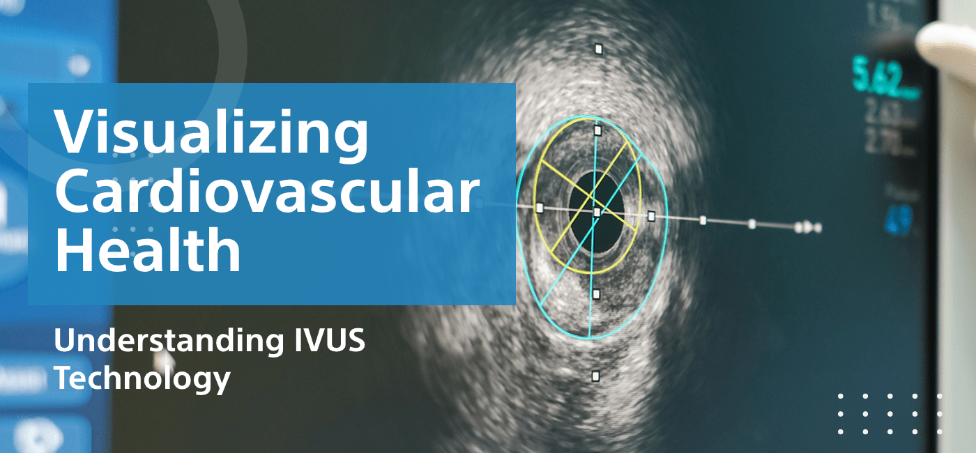
The generally utilised ultrasound imaging through precision depends on a device that is in the form of a catheter, which is inserted into the coronary vessels. While the catheter is pushed through the blood vessel, the ultrasound waves reflect from the walls, signal processor which represents the blood vessel cross section. The catheter allows for the coronary conduit, which is essentially a transmission line, to be photographed in a 3D format, from the sides of the walls of the vein. IVUS not only provides exact morphological details about the degree and extension and tissue organisation and functioning path of the coronary disease which is nearly impossible to find out through angiography. It conceivably can better assess the degree of stenosis than angiography alone. These small details contribute greatly to the detection of CAD, making it easier for specialists to diagnose, detect and treat the disease.
The IVUS value of stent placement demonstrates the crucial impact of the technology
Restoring coronary route flows via methods of coronary stent implantation is a conventional method for increasing blood stream in patients’ coronary illness. To sum up, CT angiography might be the only means that can compare to the traditional angiography as far as it concerns the size and positioning of the stent. IVUS is the basis for a rational and correct stent placement algorithm. The pre-stenting IVUS assessment plays a crucial role in determining exact size and length of the stent, based on the severity of the disease, and in determining the exact area for stent placement. The screen following the stenting area proves to be more helpful as it gives us a better view of apposition of stent to the vessel wall as well as angiography and stenosis. While the IVUS as a flaw detector empowers specialists to quickly actualize the necessary remedial measures and minimise complexities, the need for further testing is not negated. This way IVUS shows its importance for the speeding of the process of percutaneous coronary interventions. This quickly resolves the issue during the complex coronary illnesses when the main trunk or the bifurcation is left.
In addition, the utility of IVUS imaging has been widely recognized in numerous other interventions like structural heart disease, cardiac toxicity, and cancer.
Apart from presenting the preference of stent placement, IVUS is also commonly used in various coronary interventions. It is given the direction of balloon angioplasty methods regarding facilitation and dissemination. IVUS is used, not only for the analysis of issues like incredibly high calcium levels in the arteries, which result in luminal stenosis, but also to direct these effective rotational atherectomy or orbital atherectomy systems. IVUS, next, is important in vain graft diseases evaluations as a tool for seeing the flow that helps to prevent rerun. It is, ultimately, IVUS that acts as an auxiliary device in all the angiography research centre strategies.
Advancements Refining IVUS Capacities
Innovation, which is on its way to fine-tuning the IVUS capabilities, strives for better results. The more recent additions such as iMAP-IVUS (Integrated Medtronic Assessment of Physiology), which combines both the evaluation of plaque anatomy and functional measurements like intracoronary pressures, are some of the latest technologies that contribute to the minimally invasive treatment of cardiac diseases. This is information on the disease’s severity and how it progresses over time. This ensures that healthcare is more effective and up to date. The newly photonics-based IVUS approach and techniques are expected to help resolve some of the issues associated with standard IVUS. The investigation was done to discern whether deployment of IVUS together with other imaging approaches like OCT (Optical Coherence Tomography), NIRS (Near Infrared Spectroscopy), pressure wire assessment etc. can further enhance the IVUS capabilities in terms of improved imaging and patient management of cardiovascular diseases.
The panoramic perspective that the EHR system provides is the shaping factor of patient care.
Thus, IVUS is becoming less new but still a fast-growing imaging tool that makes possible the more wide-spread and outside-inside visualisation of cardiovascular infection. Through the surgical details obtained by Intravascular Ultrasound imaging, the physicians can thoroughly analyse the disease process and plan to tailor individual treatment for complex interventions as well as assess its severity and find out the areas of difficulties. Such a beautiful view will be instrumental in the patient outcome development, not only by enabling precise analysis and planning but also by incorporation and putting into action diverse strategies for treating cardiovascular disease effectively. This innovation, IVUS, undeniably will, and may be the platform that will determine the future of cardiovascular care.